![]() Views: 39
Views: 39
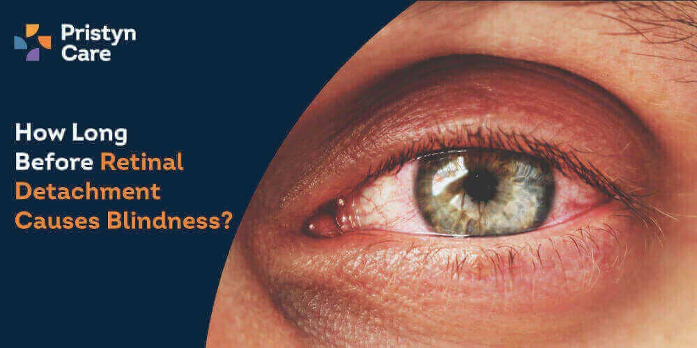
How Long Before Retinal Detachment Causes Blindness?
The causes of retinal detachment often relate to age, trauma, or other eye conditions, but the speed at which vision deteriorates can vary significantly based on the severity and location of the detachment.
Dedicated Support at Every Step!
Our Doctors are available 24 hours a day, 7 days a week to help you!
Call Us9513-316-643The key to preserving vision is timely intervention, typically through retinal detachment surgery, which can repair the detachment if performed before the retina is irreversibly damaged.
This discussion will help you know about the urgent nature of this condition, emphasizing why immediate medical attention is crucial for preventing the potentially rapid progression toward blindness.
Table of Contents
What is Retinal Detachment?
Retinal detachment is a serious ocular condition where the retina, the thin layer of tissue that lines the back of the eye, becomes separated from its underlying support tissue. This separation prevents the retina from functioning correctly, leading to potential vision loss or blindness if not treated promptly.
There are three main types of retinal detachment: rhegmatogenous, the most common type, occurs due to a break or tear in the retina that allows fluid to accumulate underneath; tractional, which results from scar tissue pulling the retina from its normal position; and exudative, caused by fluid leakage without a tear.
Symptoms of retinal detachment often develop rapidly and can include flashes of light, floating spots, or a shadow veiling the side or middle of the visual field, which may signify the retina beginning to pull away from its supportive tissue. Early detection of these warning signs is crucial for effective treatment.
No Cost EMI, Hassle-free Insurance Approval
How Retinal Detachment Occurs?
Retinal detachment is a critical eye condition that happens when the retina, the thin layer of nerve tissue lining the back of the eye, becomes separated from the underlying supportive tissue. This detachment disrupts the retina's ability to process visual signals, leading to potential vision loss.
Typically, the condition starts with a tear or hole in the retina, which allows fluid from the vitreous gel that fills the eye to pass through and accumulate under the retina, lifting it away from its normal position.
Effects on the Eye and Vision
Once the retina detaches, it is deprived of crucial nutrients and oxygen supplied by the blood vessels in the underlying tissue. This deprivation can rapidly lead to the deterioration of photoreceptor cells, which are essential for vision. Initially, symptoms might include the appearance of floaters and flashes of light.
As the condition progresses, a dark shadow may appear to advance across the visual field, starting peripherally and moving centrally, leading to significant vision loss if untreated.
Critical Timelines and Risk Factors
The urgency of treating retinal detachment cannot be overstated, as the timing of intervention greatly affects the prognosis. Key factors that influence the urgency include:
- Location of the detachment: Detachments involving the macula, the central part of the retina responsible for detailed vision, require immediate attention.
- Size of the detachment: Larger detachments cover more of the retina and pose a higher risk of extensive vision loss.
- Underlying health conditions: Patients with diabetes or other vascular conditions may experience more rapid progression.
- Previous ocular history: Eyes that have undergone surgeries or trauma may have different risk profiles and timelines.
Factors Influencing the Speed of Vision Loss
- Extent of the detachment: The larger the area of the retina affected, the greater the potential for vision loss.
- Involvement of the macula: If the macula is detached, vision loss can be more severe and immediate.
- Duration before treatment: The longer the retina remains detached, the higher the risk of irreversible damage.
- Patient’s overall eye health: Pre-existing conditions such as diabetic retinopathy can exacerbate the effects of a detachment.
- Age: Older patients often experience more severe outcomes due to the less resilient nature of their retinal tissue.
Timeline from Retinal Detachment to Potential Vision Loss
The timeline for potential vision loss following retinal detachment can vary. In cases where the macula is not initially affected, patients might notice a gradual loss of peripheral vision. However, if the macula detaches, central vision can deteriorate within hours or days, emphasizing the need for urgent retinal detachment surgery.
Diagnosis and Detection
Early diagnosis is crucial for successful treatment outcomes. The best ophthalmologist will employ a variety of diagnostic tools to confirm the presence of retinal detachment. These may include:
- Dilated eye examination: Essential for visualizing the retina directly.
- Ultrasound imaging: Used if the retina is not visible due to opacities in the eye.
- Optical coherence tomography (OCT): Provides detailed images of the retinal structure to assess the extent of detachment.
Accurate and early diagnosis not only facilitates immediate treatment but also significantly improves the chances of recovering vision. As such, anyone experiencing symptoms of retinal detachment should seek medical advice immediately to prevent permanent vision loss.
Understanding the causes of retinal detachment, the effects on the eye, and the importance of critical timelines can empower patients to act promptly.
MS, DNB, FICO, MRCS, Fellow Paediatric Opth and StrabismusMobile
FREEConsultation Fee
Diagnostic Methods for Retinal Detachment
Retinal detachment is a serious eye condition that necessitates prompt and accurate diagnosis to prevent irreversible vision loss. The diagnostic process typically involves several key methodologies:
- Ophthalmoscopy: This tool allows eye specialists to view the retina directly, identifying any tears or detachments.
- Ultrasound imaging: When the retina cannot be seen directly due to opacities in the eye, ultrasound provides a clear picture of the retinal position.
- Optical coherence tomography (OCT): This non-invasive imaging test gives cross-sectional images of the retina, helping detect even subtle detachments.
Role of Timely Diagnosis in Preventing Blindness
Timely diagnosis plays a crucial role in preventing blindness from retinal detachment. Detecting the condition early increases the chances of successful treatment options, allowing interventions before the retina suffers extensive damage.
It is pivotal that individuals experiencing symptoms such as sudden visual disturbances or shadows in their vision consult the best ophthalmologist immediately.
Treatment Options
Treatment options for retinal detachment are primarily surgical, aimed at reattaching the retina to its proper place at the back of the eye. The choice of procedure depends on the type and severity of the detachment:
- Pneumatic retinopexy: This involves injecting a gas bubble into the eye to push the retina back into place.
- Scleral buckle: A flexible band is placed around the eye to gently push the walls of the eye against the detached retina.
- Vitrectomy: Removing the vitreous gel and replacing it with a gas bubble or silicone oil to hold the retina in place.
Immediate Treatments and Their Effectiveness
Immediate treatments such as pneumatic retinopexy and scleral buckling are often very effective if performed soon after detachment occurs. These methods can effectively reattach the retina and restore vision, particularly in cases where the macula (the central part of the retina) is not yet affected.
Long-term Management and Surgical Options
Long-term management may involve vitrectomy, especially for severe detachments or proliferative vitreoretinopathy (PVR), a complication that can arise after initial treatment. Ongoing follow-ups are essential to monitor the eye's health and the attachment of the retina.
Prognosis After Retinal Detachment
The prognosis after retinal detachment varies widely. Factors influencing the outcome include the extent of detachment, the involvement of the macula, and the timeliness of intervention. The recovery process can take several months, during which vision may gradually improve.
Recovery Rates and Success Statistics
The success rate of retinal detachment surgery can be high, with about 80-90% of cases successfully repaired with a single operation. However, the overall visual recovery varies. If the macula was detached, the visual outcome might not be as good compared to cases where the macula was not affected.
Long-term Visual Outcomes
Long-term visual outcomes depend significantly on the initial condition of the retina and the macula at the time of the surgery. Regular monitoring and management can help maximise visual recovery and reduce the risk of future detachments.
Prevention and Monitoring
Effective preventions involve managing risk factors such as controlling diabetes, protecting the eyes from trauma, and monitoring for eye diseases that could predispose one to retinal detachment. Regular eye exams are essential for early detection and effective prevention of serious complications.
Preventative Measures and Regular Eye Exams
Regular eye exams are critical for detecting changes in the retina that may signify the early stages of detachment. These exams are especially important for individuals with high myopia, previous eye surgeries, or a family history of retinal detachment.
Monitoring Techniques for At-Risk Individuals
For those at higher risk, more frequent and detailed eye examinations may be recommended. Techniques such as peripheral vision testing and regular OCT scans can help detect early signs of retinal tears before they progress to full detachment, enhancing the effectiveness of preventions.
Through comprehensive care, including effective treatment options, regular monitoring, and adherence to preventive measures, individuals can maintain optimal eye health and reduce the risk of serious visual impairment due to retinal detachment.
Conclusion
Retinal detachment can swiftly lead to significant vision loss, highlighting the critical need for swift action. If the macula is affected, the window to preserve sight narrows dramatically. Prompt
Retinal Detachment surgery is vital for stopping further damage and enhancing the chances of vision recovery. Regular eye check-ups and immediate response to symptoms are essential to effectively safeguard one's vision against the rapid onset of blindness due to retinal detachment.


