Diagnosis
The doctor asks about the symptoms and carries out a thorough physical exam to look for areas of swelling, discolored skin, and soreness. If the doctor suspects the presence of an underlying blot clot, they ask for certain tests.
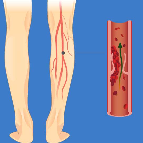
Deep vein thrombosis is usually diagnosed through a Doppler’s ultrasound. In Doppler’s ultrasound, high-frequency sound waves are used to check the flow of blood through the veins. The ultrasound detects the blood flow and an impaired blood flow indicates the presence of blood clots in the vein.
The doctor may conduct a series of ultrasounds to check if the blood clot is growing or to look for new blood clots that may have formed.
Surgery
The doctor asks about the symptoms and carries out a thorough physical exam to look for areas of swelling, discolored skin, and soreness. If the doctor suspects the presence of an underlying blot clot, they ask for certain tests.
Deep vein thrombosis is usually diagnosed through a Doppler’s ultrasound. In Doppler’s ultrasound, high-frequency sound waves are used to check the flow of blood through the veins. The ultrasound detects the blood flow and an impaired blood flow indicates the presence of blood clots in the vein.
The doctor may conduct a series of ultrasounds to check if the blood clot is growing or to look for new blood clots that may have formed.
In the beginning, the doctor will provide medications (blood thinners like heparin, warfarin, enoxaparin, or fondaparinux) or suggest using compression stockings to improve the blood flow and minimize the chances of blood clot formation.
If medical treatment doesn’t work, the doctor will recommend other methods. Deep vein thrombosis is most commonly done in combination with one or more of the following treatment methods:
- Thrombolysis
- Anticoagulant medications
- Angioplasty & stenting
- Thrombectomy
- Placing a vena cava filter in the deep vein.
Thrombolysis- It is also known as thrombolytic therapy or fibrinolytic therapy in which certain medications are used to break the blood clots that are formed in the vessels. The most common medicine that is used in this technique is tissue plasminogen activator (tPA) but there are other medicines also available.
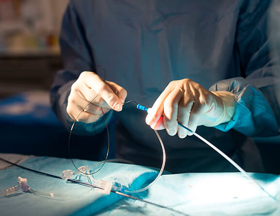
IVC (Inferior Vena Cava) Filter- The IVC filter is a metal device that looks like an umbrella and can stop the blood clot’s movements. It is placed inside the main vein, i.e., the inferior vena cava that runs through the belly. An incision is made around the abdomen and a catheter is inserted into the vein using an X-ray guide. The filter is placed over the blood clot inside the vein and it attaches itself to the walls of the vein.
Thrombectomy- Also known as venous thrombectomy, it is a procedure that involves surgical removal of the vein clot. During thrombectomy, the vascular surgeon will make an incision into the blood vessel to remove the blood clot. Then the blood vessels and tissues are also repaired.
Angioplasty- In some cases, angioplasty or the balloon suction technique may be used to keep the vein inflated and a stent may be used to hold it open while the blood clot is being removed. When the blood clot is extracted, the balloon is also taken out simultaneously.
The surgical methods available for DVT treatment are not without risks. Therefore, you should always make sure that you are getting treatment from the best vascular specialist in Kolkata.


.svg)


